$ 44275
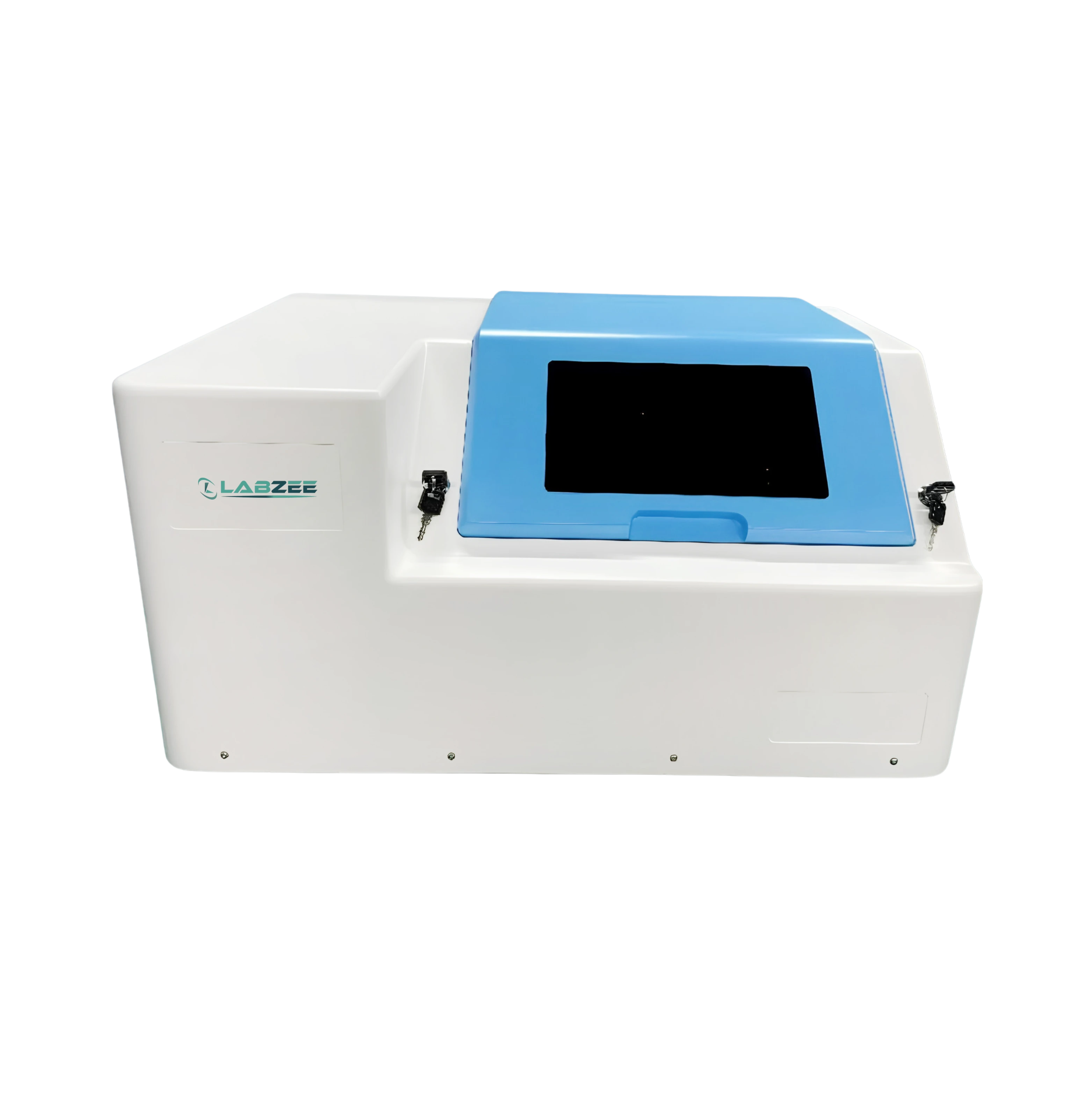
| Camera Resolution | 3200×2200, 1” global shutter |
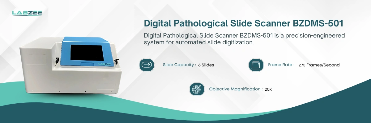
| Slide Capacity | 6 Slides |
| Frame Rate | ≥75 Frames/Second |
| Objective Magnification | 20x |
| Numerical Aperture (NA) | 0.75 |
| Scan Speed | ≤180 Sec/Sample |
| XY Driver Resolution | 5 μm |
| Z Auto Focus Resolution | 0.25 μm |
| Image Resolution | ≤0.25 μm/Pixel |
| Light Source | LED, >20,000 Hours |
| Scan Operation | One-Click Scan, Pause/Stop, Sample Add-In |
| Image Format | JPEG, BMP |
| PC Configuration | Embedded PC, NVIDIA GPU, keyboard, USB 3.0 |
| Software Features | Auto Smear Contour Detection, Targeted Scan |
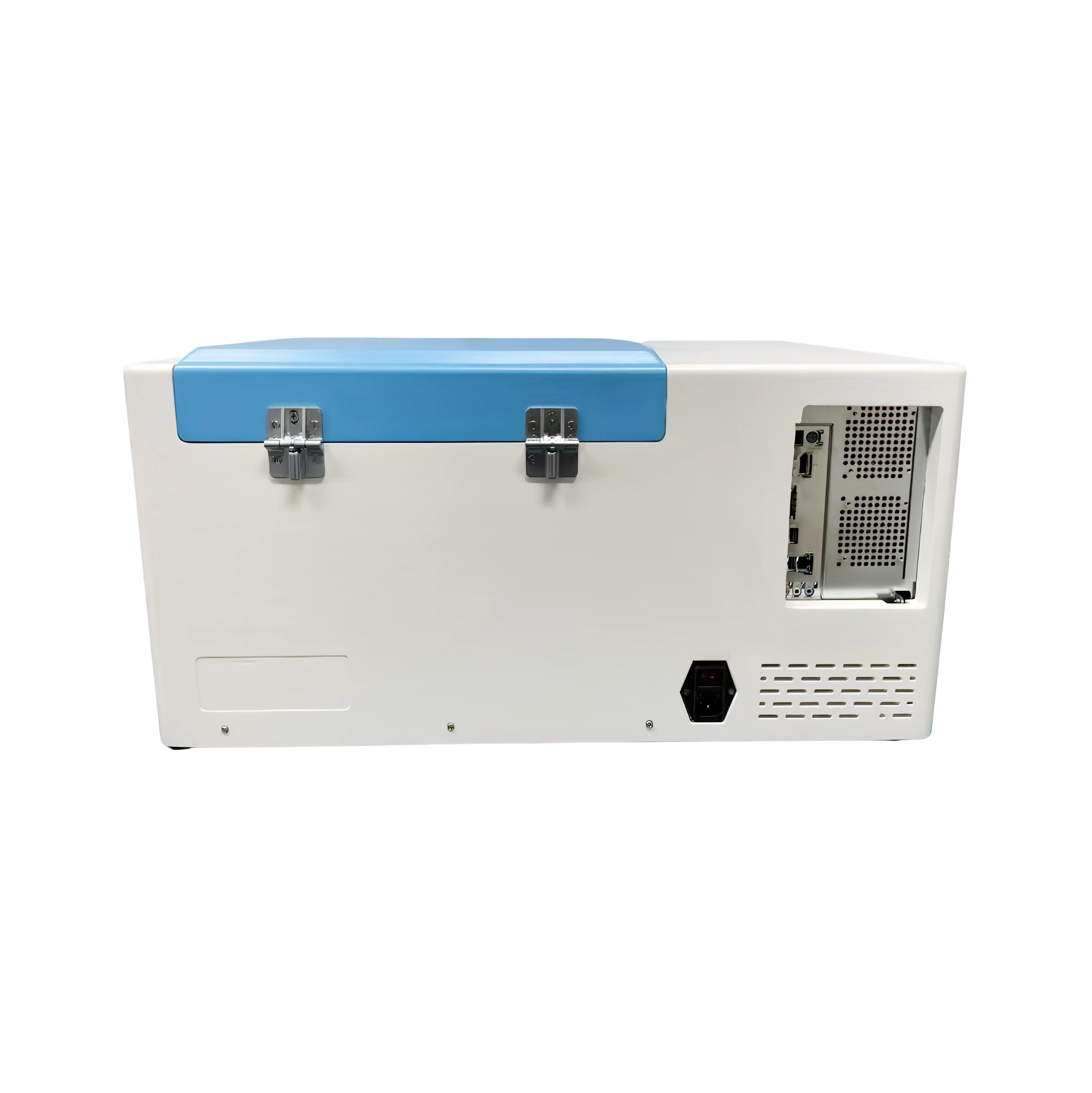
| Product Dimension | 698 × 464 × 345 mm |
| Packing Dimension | 800 × 550 × 550 mm |
| Net Weight | 35 Kg |
| Gross Weight | 75 Kg |
• Auto Smear Contour Detection: Detects smear contours for targeted image acquisition.
• Targeted Scanning: Scans selected areas of slides for focused image capture.
• One-Click Scan Operation: Starts the scanning process with a single command.
• Pause/Stop Function: Allows scanning to be paused or stopped during operation.
• Sample Add-In Function: Allows additional slides to be added during the scanning process.
• Whole-Slide Imaging: Captures digital images across the required slide area.
• Image Resolution: Supports detailed digital slide imaging for pathology review.
• XY Stage: Supports controlled slide movement during the scanning process.
• JPEG and BMP Image Formats: Supports commonly used formats for digital image viewing and documentation.
• LED Light Source: Provides illumination for digital slide scanning.
• Pathology Laboratories: Supports tissue slide examination and documentation.
• Histology Laboratories: Assists with tissue section review and record keeping.
• Clinical Research: Covers microscopic sample evaluation during research studies.
• Telepathology: Enables remote sharing and review of pathology slides.
• Pharmaceutical Research: Enables laboratory sample examination and research activities.
Frequently Asked Questions
1: What camera technology does Digital Slide Scanner BZDMS-501 use?
Digital Slide Scanner BZDMS-501 uses a global shutter camera system that ensures high-quality, motion-free imaging, enabling clear scans even at fast frame capture speeds for dynamic pathology environments.
2: Which scanning features enhance automation in BZDMS-501?
The scanner includes advanced features like one-click scanning, pause or stop functions, and sample add-in capability, streamlining workflow without manual interruption.
3: Can you explain how Digital Slide Scanner BZDMS-501 supports smear analysis?
The scanner’s software includes auto smear contour detection and targeted scan capabilities that help isolate specific areas of interest on slides, optimizing both accuracy and efficiency during digital smear reviews.
4: How does BZDMS-501 maintain scanning accuracy?
It maintains high scanning precision through autofocus resolution and XY stage control, ensuring consistent focus and movement accuracy, which are crucial for digital pathology applications.
5: What makes Digital Slide Scanner BZDMS-501 suitable for high-throughput use?
With multi-slide capacity, integrated PC with GPU, and fast scanning speed, the BZDMS-501 is built to support continuous, high-volume digital imaging demands in clinical diagnostics and research labs.
6: When is a digital slide scanner used in pathology labs?
It is used to digitize stained tissue slides for viewing, analysis, and sharing. It enhances archiving, remote diagnostics, and integration with AI tools. Digital scanning supports faster, collaborative pathology workflows.
7: Can multiple slides be scanned in a single run?
Yes, most models support batch loading and scanning of dozens to hundreds of slides. Automated feeders help manage large volumes efficiently. This is ideal for high-throughput environments.
8: Can scanned slides be integrated with pathology software?
Yes, scanners are designed for compatibility with digital pathology platforms and LIS. They allow seamless image storage, annotation, and case management. Some models support cloud-based access and AI analysis.
9: When should calibration be performed for consistent image quality?
Regular calibration-typically monthly or as recommended-is vital for optimal image clarity. Some systems include auto-calibration features. Proper maintenance ensures diagnostic reliability.
10: Can different magnifications be selected during scanning?
Yes, users can choose from various magnification levels (e.g., 20x, 40x) based on diagnostic need. Dual or multi-objective systems provide flexibility. High magnification improves detail but may increase scan time.
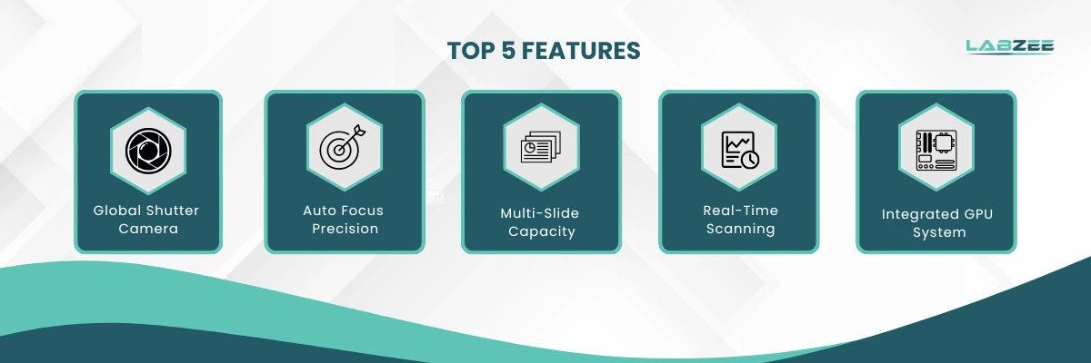
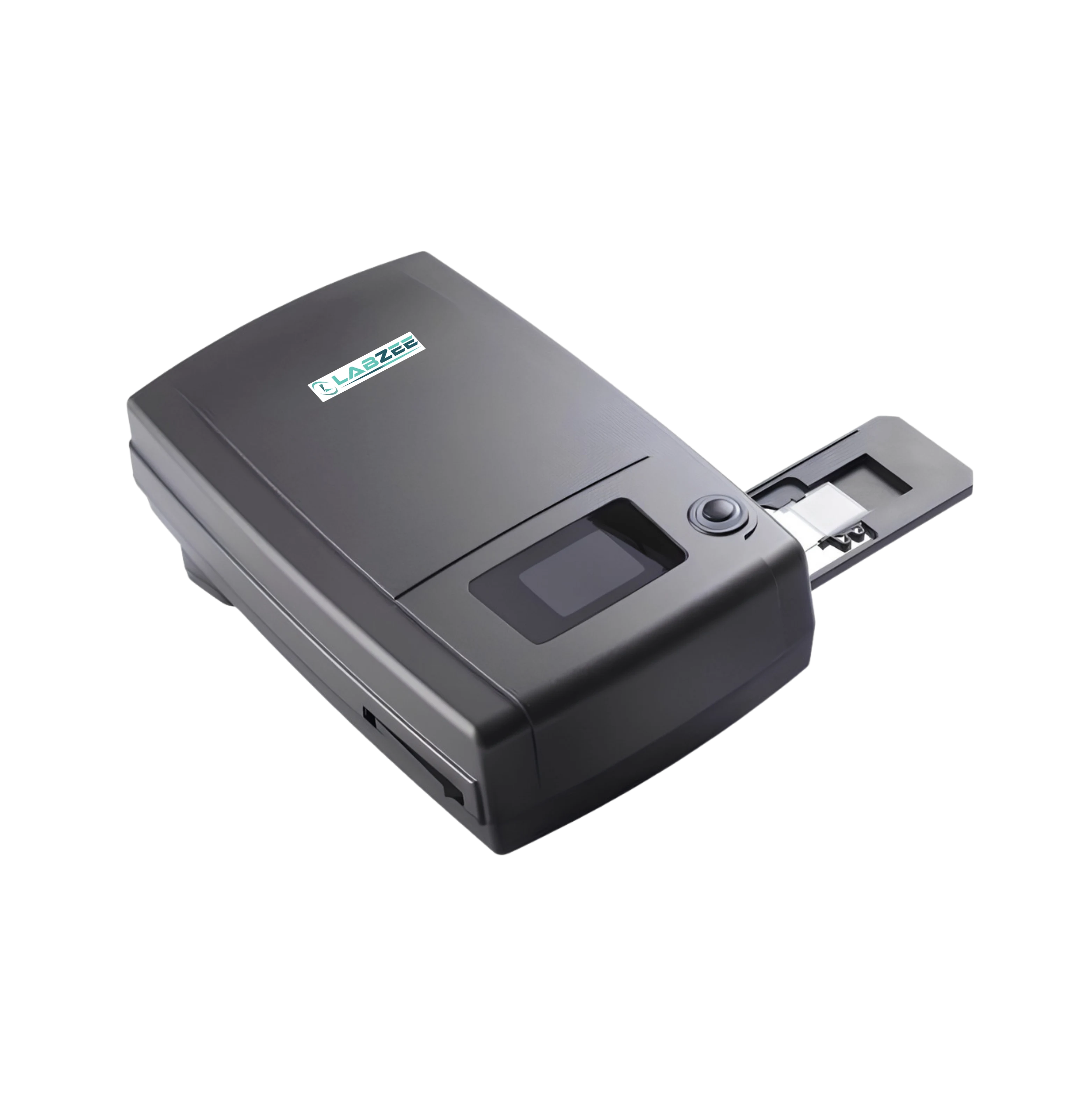
Digital Pathological Slide Scanners from Labzee offer imaging instruments used to convert traditional glass microscope slides into high-resolution digital images for viewing, analysis, documentation, and sharing. Digital Pathological Slide Scanner features support Digital Pathology Imaging through Automated Image Acquisition with a Global Shutter Camera for image capture. Digital pathology scanners support Automated Slide Scanning, multiple slide capacities, Whole-Slide Imaging, and different Objective Magnification options for pathology and histology workflows. Automated Digital Pathological Slide Scanners help process multiple slides in a single scanning run through Multi-Slide Scanning, with configurations available for 6, 60, 120, and 240 slides, along with additional capacity options. Digital pathology whole-slide scanners support detailed image analysis and digital slide review using Z-Axis Autofocus and an XY Stage for slide positioning during scanning.
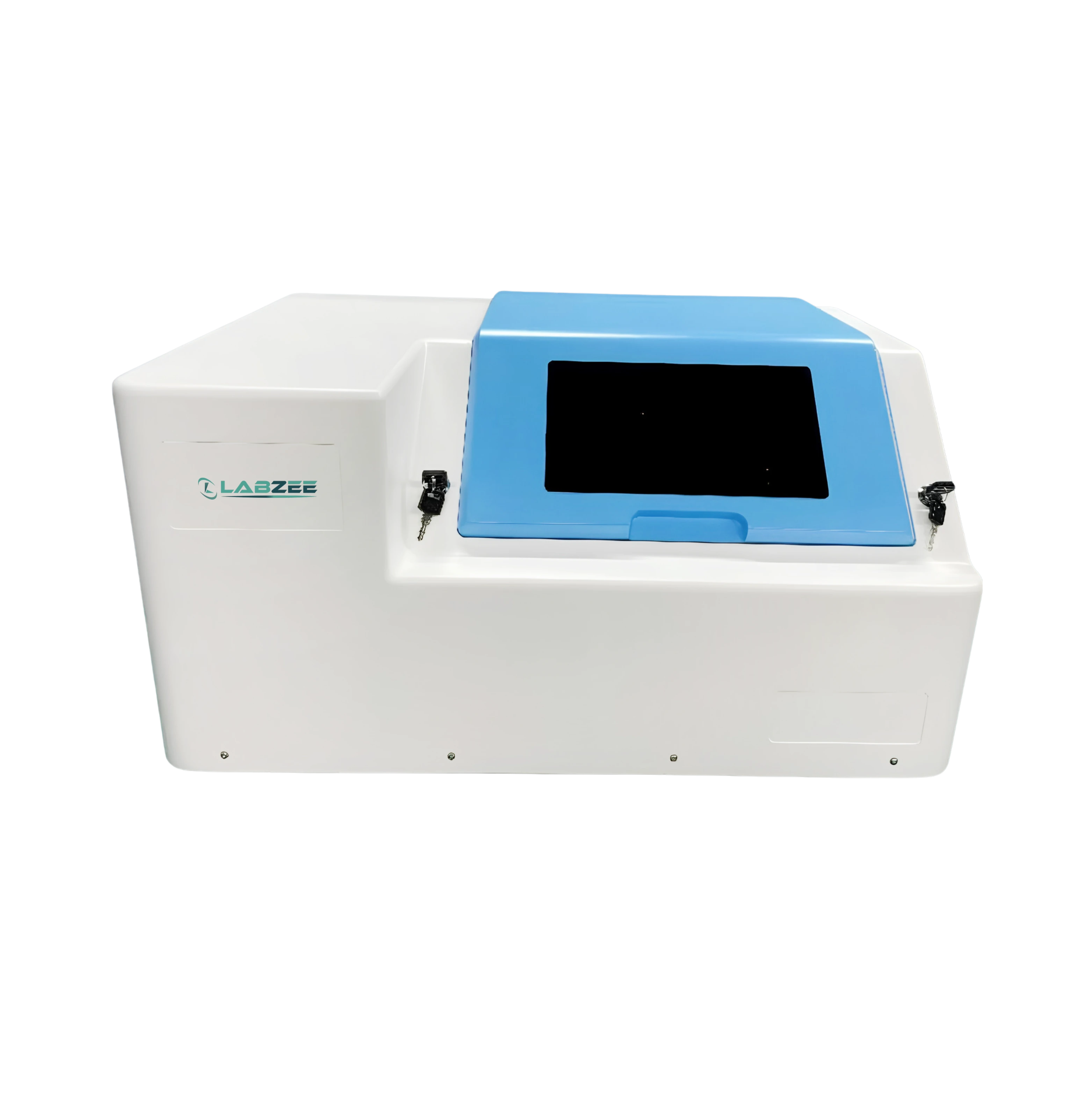
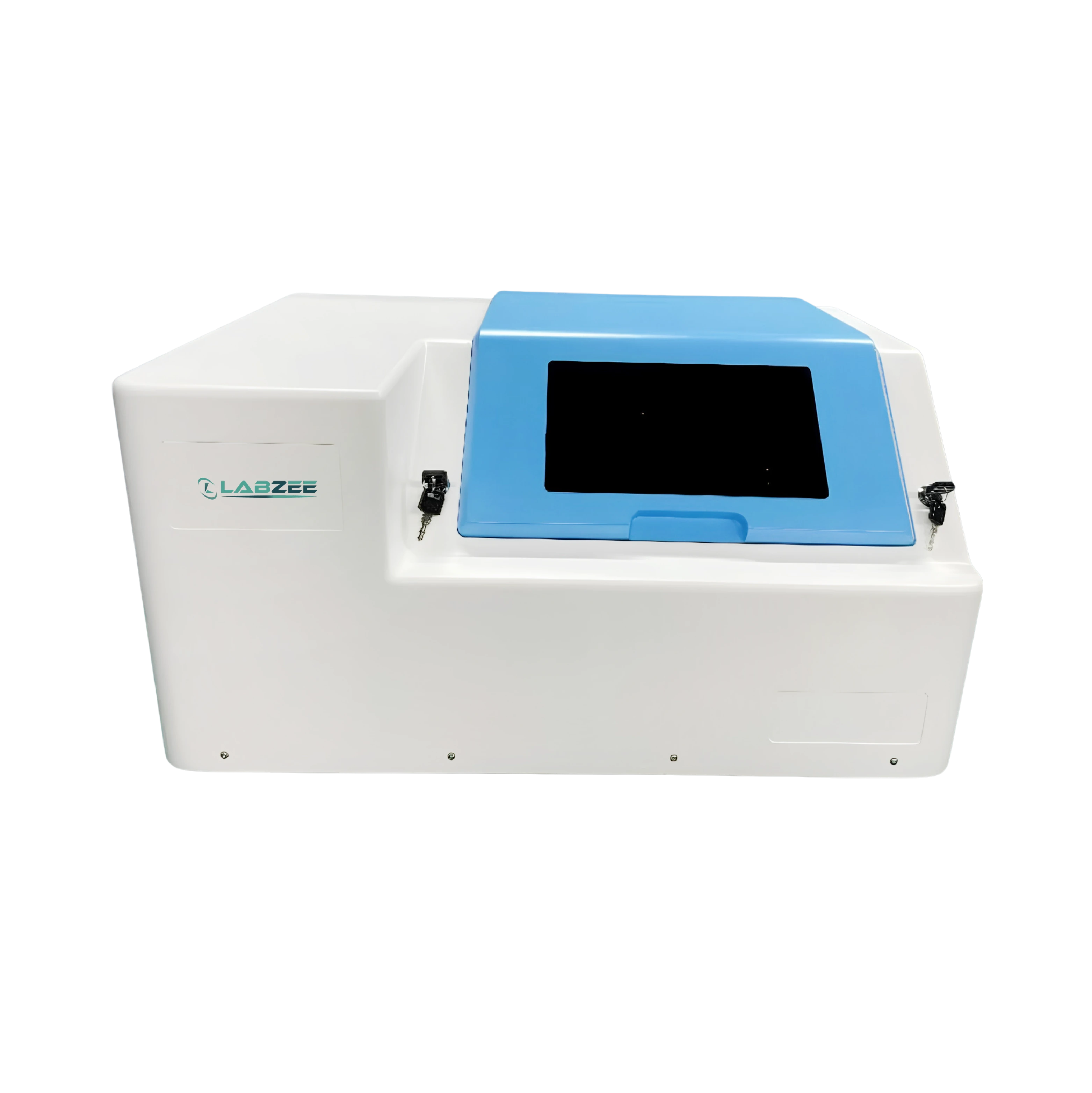
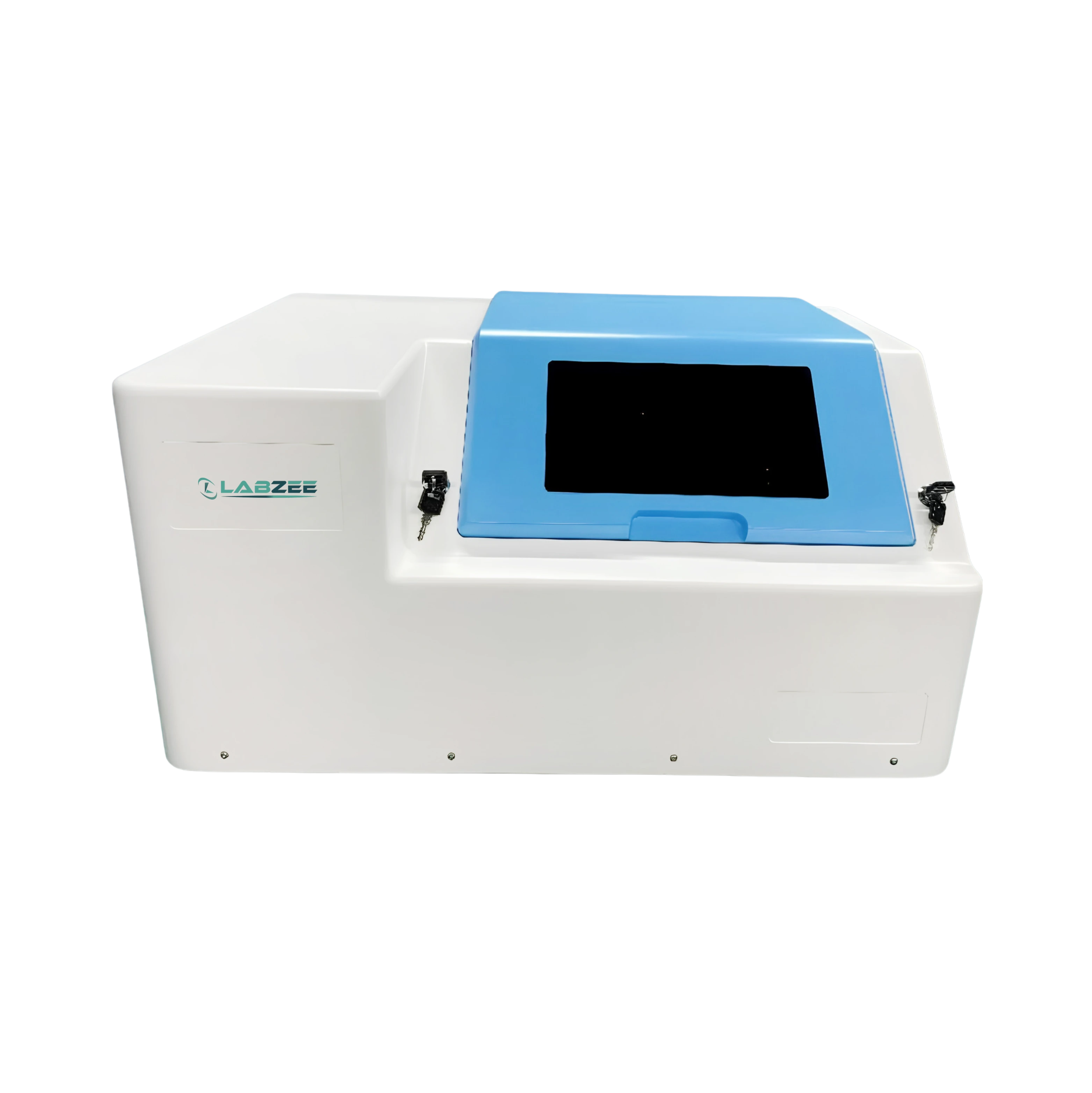
Pathological Slide Scanners from Labzee provide digital imaging of microscope slides for pathology and histology workflows, covering tissue-slide digitization, image review, and documentation. Digital pathology scanners perform automated slide scanning and whole-slide digitization for pathology and histology workflows. Our scanners combine optical magnification, image resolution, dynamic range, and automated slide handling for detailed digital slide imaging to support tissue-section examination and documentation.
$ 4078
